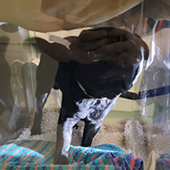
Pictured: Gary inside the fluoroscopy container.
The unique box allows for safer and easier procedures.
Veterinary surgeons at the University of Edinburgh's Hospital for Small Animals have designed a tool to make fluoroscopy procedures safer and easier to conduct.
Fluoroscopy, a non-invasive imaging technique, can be very difficult to conduct, as the patients must stand still on their own, to avoid the veterinary team being exposed to the electromagnetic waves used.
The specialist Internal Medicine Service team at the Hospital for Small Animals designed and developed a solution to keeping the patients still enough to conduct a fluoroscopy procedure – a clear acrylic box, which patients can stand in during swallow studies.
With no metal components, the container allows for clear and effective images.
The effectiveness of the fluoroscopy container was proven when a three-year-old pug named Gary was referred to the hospital while suffering from sever abdominal cramping. With the help of the fluoroscopy container, the veterinary team was able to conduct some swallow studies and diagnose some acid reflux.
Dr Silke Salavati, senior lecturer in Small Animal Medicine, commented: “Due to the availability of the live fluoroscopy and other specialist imaging techniques, our specialist diagnostic imaging teams can pick up subtle changes like the acid reflux in Gary’s case, which helps to optimise treatment.”
Images (C) The University of Edinburgh



 Birmingham Dogs Home has issued an urgent winter appeal as it faces more challenges over the Christmas period.