Puppies with spina bifida treated with combination of surgery and stem cells
Placental stem cells were developed by researchers at UC Davis School of Medicine, then characterised and combined with a tissue-engineered scaffolding to optimise treatment by UC Davis veterinary surgeons.
Dogs born with spina bifida frequently have little control of their hindquarters and are typically euthanised as puppies. This latest research offers hope for similar cases in the future, as well as a basis to inform human clinical trials for babies with the birth defect.
The surgical techniques, developed by Dr Diana Farmer, professor and chief of surgery at the UC Davis, combined with the stem cell treatment developed by the research faculty, are progressions toward her goal of curing spina bifida.
She has already pioneered the use of surgery prior to birth to improve brain development in children with spina bifida; and that prenatal surgery – combined with placenta-derived mesenchymal stromal cells (PMSCs), held in place with a cellular scaffold – preserved the ability of lambs born with the disorder to walk without noticeable disability.
Dr Beverly Sturges, professor of neurosurgery at the UC Davis veterinary hospital, wanted to find out if Dr Farmer’s surgery-plus-stem-cell approach could give dogs closer-to-normal lives, along with better chances of survival and adoption.
So, 10-week-old, bulldog siblings, Darla and Spanky, were transported from the Southern California Bulldog Rescue to UC Davis. After a few days of monitoring to see that they were healthy enough for the procedure, Darla underwent surgery on the third day of hospitalisation and Spanky on the fourth.
In surgery, a myelomeningocele was identified at the L7 region of both dogs, consistent with the spina bifida identification on their MRIs. The dogs displayed dorsal positions of the lower extremities of their spinal cords, which were tethered. The sites were dissected and replaced in their spinal canals, with stem cells being surgically placed over the defects.
The disorder usually becomes apparent between one and two weeks of age, when puppies show hind-end weakness, poor muscle tone, incoordination, incontinence and abnormal use of their tails. At their two-month recheck appointment, Darla and Spanky – although still incontinent – showed off their abilities to walk, run, and play and both dogs seemed to have an improved range of motion and control of their limbs.
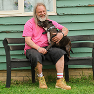
The dogs have since been adopted, and continue to do well at their new home in New Mexico.
Image (C) UC Davis.